Skip to:

MRFN Member Login
Program Application

Intermediate Small Angle Diffractometer (2-Circle)

Instrument types

Features

  • X-ray source: Rigaku UltraX18 rotating anode (Cu anode, wavelength = 1.54 Å)
  • Monochromator:Osmic Confocal multilayer mirror
  • Beam size at sample position: ~0.8 mm x 0.8 mm
  • Detector: Mar180 (18 cm diameter)image plate detector
  • Optics: 2 sets of motorized Huber slits with newly developed scatterless hybrid slit blades
  • Flight paths: evacuated both on the incident and diffracted sides
  • Goniometer: large open Huber 2-circle
  • Sample stage: motorized X-Y-Z-THETA for transmissiona and grazing-incidence SAXS
  • Automated sample changing: up to 14 x-ray capillaries with full x-y-z positioning for each sample
  • Filter inserter: 3 filters and 1 shutter
  • Flux: ~ 109photons/second
  • Typical range of lengthscale probed: 10 Å – 200 Å
  • Speciment-to-detector distance: typically 750 mm
  • Computer:Linux PC
  • Software for data collection: SPEC
  • Software for data processing: MARVIP and FIT2D
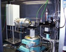
    In-situ GI-SAXS data of an ordered sol-gel grown non-silicate oxide film (Boettcher 2007) SAXS data of a mesostructured silica film with a bicontinuous cubic structure (Hayward 2004)